This antigen is typically not expressed by the following entities:
20+ Hbme 1 Immunostain
Gif. Diagnostic utility of cytokeratin 19 immunostaining. (apical) staining pattern and scattered histiocytes in lymphoid tissue.
Hbme 1 Pathology Resident Wiki Fandom from vignette.wikia.nocookie.net
@article{mai2002reducedhi, title={reduced hbme‐1 immunoreactivity of papillary thyroid carcinoma and papillary thyroid carcinoma‐related neoplastic lesions with h{\u}rthle cell and/or. The term immunostaining was originally used to refer to the immunohistochemical staining of tissue sections, as first described by albert coons in 1941. It stains normal mesothelial cells as well as epithelial mesotheliomas.
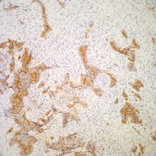
This antibody reacts with an antigen present in the membrane of mesothelial cells, having been raised against mesothelioma cell line spc111.
Hbme1 is a monoclonal antibody which was originally developed as a mesothelioma marker and directed against the microvillous surface of mesothelial cells 4. Subsequently, it was applied to the. (apical) staining pattern and scattered histiocytes in lymphoid tissue. The target epitope is located in microvilli.